

[In the summer of 2026, I had the opportunity to conduct research in the Khare Laboratory at Rutgers University under the mentorship of Dr. Sagar Khare and Austin Seamann. The Khare Lab is part of the Institute for Quantitative Biomedicine (IQB), and Dr. Khare is a Professor in the Department of Chemistry and Chemical Biology. My project focused on using AI-based protein design tools to design a binder for a photoswitchable protein. Below is a summary of the research, methods, and key outcomes from the project. ]
Designing a binder for a photoswitchable protein
Hello! This is the first in a series of posts that will cover my summer research project on molecular engineering. The overall system we are looking to construct aims to support the synthesis of single-stranded DNA without a template (Fig. 1). Its is an ambitious project, requiring the collaboration of many. In this series of posts, I have focused on one step of the process: designing a new protein binder for Cph1, a photoswitchable protein.
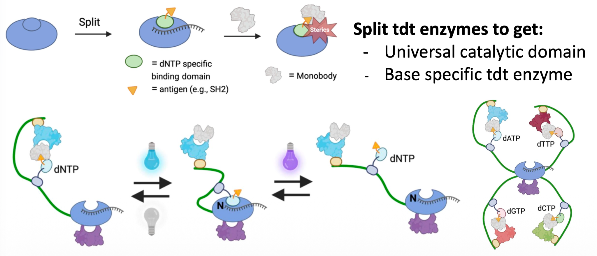
Why a protein like that? you may ask. First of all, we need to look at the overall project. The idea is to design a system that uses split TdT enzymes. These enzymes add nucleotides to the 3’ end of a DNA molecule, enabling the construction of our target product. One part of the system would act as a universal catalytic domain, while another part would be specific to a DNA base. For the system to work, we need to put together the necessary components, and that’s where Cph1 comes in handy.
A protein controlled by light
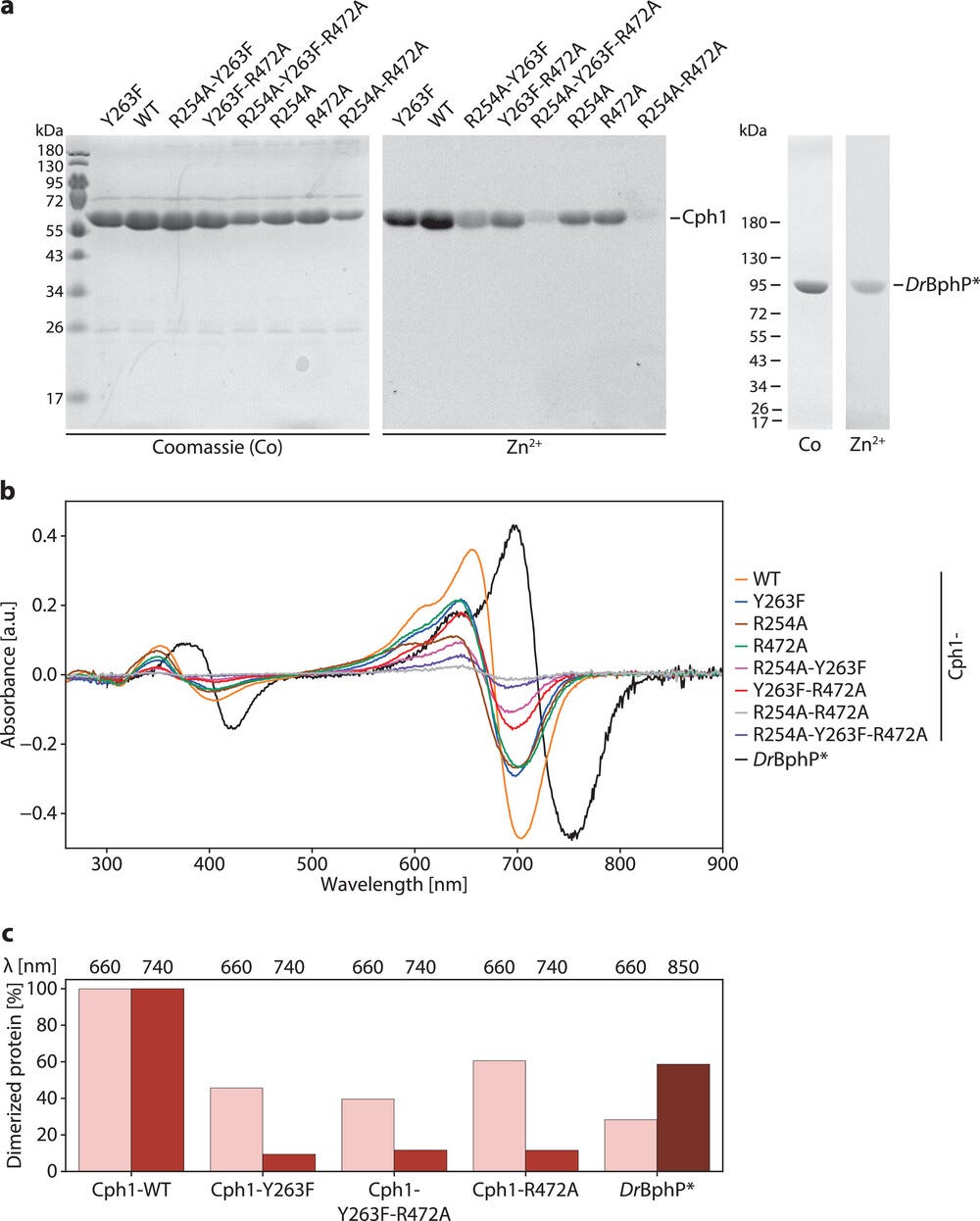
Cph1 is a photoswitchable protein. This means that we can turn the light/bright state of the protein on and off with the correct light pulse stimulus. But this not the only property of Cph1; it is also a dimer, two similar units bonded together, called Chains A and B (Fig. 2). An engineered Cph1 variant has a really cool feature: it can change between dimeric and monomeric states when exposed to different wavelengths of light. In the light state, exposure at 616 nm can promote dimerization, whereas exposure at 740 nm can return the protein to a dark state and lead to monomerization (Fig. 3). This reversible change creates a possible way to control when two protein components come together, a key step in our TdT-system.
Dimerization levels for wild-type Cph1 and engineered variants under different light conditions
Finding the location for a binder
Determining the binder location.
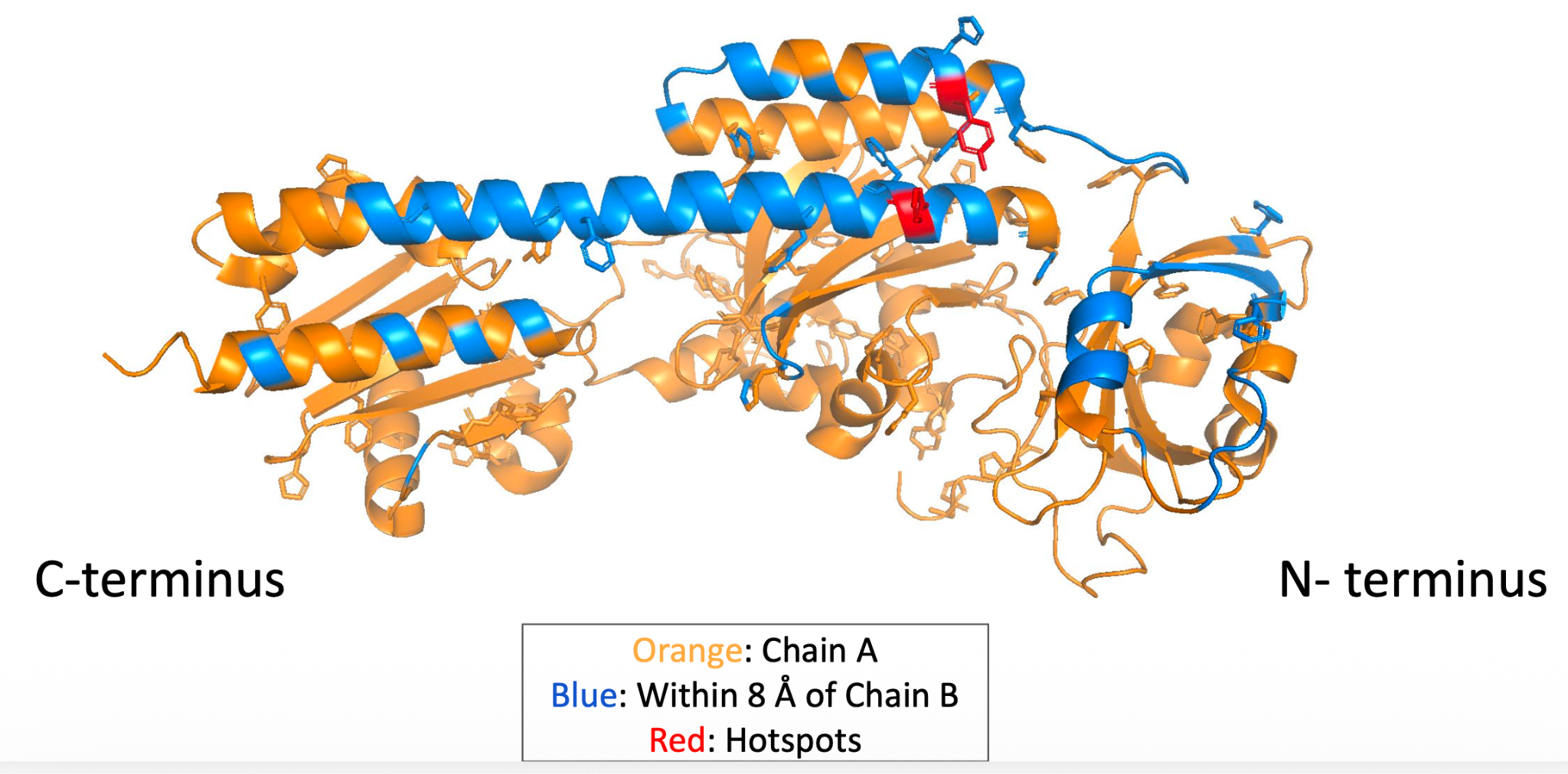
To design the binder, we first needed to decide where exactly it should interact with Cph1. The most logical step was to examine the interface where Chains A and B meet. This is the region involved in dimerization, and the goal was to place the binder near the center of this interface. We used the software PyMOL, an open-access molecular visualization software, to inspect the Cph1 structure and identify the interface. With this procedure we were able to choose the most favorable region for the binding hotspots. We selected residues A307 and A137, which were described as hydrophobic and aromatic residues (Fig. 4).
The selected binder-design region on Chain A. Blue residues lie within 8 angstroms of Chain B, and red residues mark the hotspots
A multi-stage design process
Now that we had selected where we wanted the binding hotspots to be, we needed to actually design them. For this process, we built a computational pipeline that relied on several computational and bioinformatic tools to model the binder candidate models. It is important to note that we generated several iterations of designs and models at eatch stage of the pipeline. Some designs looked promising at first but failed later tests. Others had shapes that were too extended, too flexible, or poorly positioned against the target. For that reason, the workflow followed a repeated pattern: generate designs, calculate metrics, remove weak candidates, and pass the remaining candidates into the next stage. This process was central to the entire project. The design models created possibilities, but the filtering and analysis scripts helped us decide which possibilities were worth keeping.
The pipeline consisted in these steps (Fig. 5):
1) Inspect the Cph1 structure using PyMOL for selecting the binding hotspots.
2) Generate new protein backbones using RFdiffusion3.
3) Generate amino-acid sequences using ProteinMPNN. These sequences needed to match the previously designed structures.
4) Place the newly generated sequences onto their intended backbone using threading.
5) Adjust the structure to improve its local arrangement using FastRelax.
6) Predict structures from the designed sequences and compare these predictions with the intended designs using AlphaFold3.

Why the project required repeated filtering
The project did not rely on a single model or a single score.
At each stage, the number of designs grew or changed. Some designs looked promising at first but failed later tests. Others had shapes that were too extended, too flexible, or poorly positioned against the target.
For that reason, the workflow followed a repeated pattern:
Generate designs, calculate metrics, remove weak candidates, and pass the remaining candidates into the next stage.
This process was central to the entire project. The design models created possibilities, but the filtering and analysis scripts helped us decide which possibilities were worth keeping.
Conclusion
Our starting point was a biological system controlled by light. From there, we identified the dimeric interface of Cph1 and selected a region where a new binder could interact with Chain A.
We then built a computational pipeline that connected structure inspection, backbone generation, sequence design, structure refinement, prediction, and filtering.
The next challenge was to run the first major generation stage at scale. That meant deciding how much of the target structure to provide to RFdiffusion3 and then finding a way to filter more than 1,000 generated binders.
Next in the series: How we used RFdiffusion3 to generate 1,082 protein backbones and reduced them to 370 candidates.